


Page 25 - Spring 2025 Electron
P. 25
Industry News


 Revolutionary Robot uses Ultrasound for early   From this virtual reconstruction clinicians can create
 Cancer Detection  cross-sectional images that mimic those generated
 by a standard biopsy.
 A tiny robot that can take 3D scans deep within the
 body using ultrasound has been developed by a   In testing an artificial colon was used with a
 team of engineers, scientists and clinicians from   robotically controlled external permanent magnet,
 the universities of Leeds, Glasgow and Edinburgh.  a platform previously developed at Leeds University   FURTHER
 which enables both joystick and autonomous control
 It paves the way  for a transformation in the diagnosis
 of the oloid magnetic endoscope. Navigation was
 and treatment of certain types of cancers by
 assisted by images from an embedded camera and   DEVELOPMENTS
 enabling ‘virtual biopsies’ (non-invasive scans) that
 a magnetic localisation system. The oloid magnetic
 provide immediate diagnostic data. This should then
 endoscope was 3D-printed from resin and has a   IN ROBOTICS
 enable doctors to detect, stage and potentially treat
 diameter of 21 millimetres. The robot could therefore
 lesions in a single procedure rather than requiring
 still roll, but was of a size and design that was suitable
 physical biopsies.
 for clinical applications.
 Results showed that the system could successfully
 perform controlled rolling and sweeping motions
 inside the colon, generate high-resolution 3D
 ultrasound scans for accurate diagnosis, and identify
 lesions in gastrointestinal tissue, so demonstrating its
          New Robotic Material mimics Biological
 potential for advanced medical imaging and early
          Processes
 disease detection.
 Human trials are due to commence in June 2026.  Researchers at Santa Barbara and TU Dresden have
          developed a proof-of-concept robotic material
 Pietro Valdastri, Professor and Chair in Robotics and   that mimics biological processes, allowing it to shift
 Autonomous Systems, and Director of the Science   between rigid and fluid states.
 and Technologies of Robotics in Medicine (STORM)
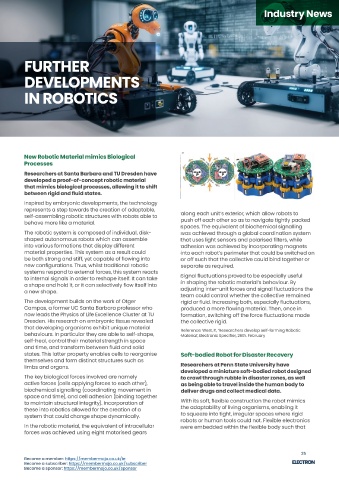
 Lab in Leeds, who also coordinated the research,   Inspired by embryonic developments, the technology
 states:  represents a step towards the creation of adaptable,   along each unit’s exterior, which allow robots to
          self-assembling robotic structures with robots able to
 “ For the first time, this research enables us to   behave more like a material.  push off each other so as to navigate tightly packed
 reconstruct a 3D ultrasound image taken from a                 spaces. The equivalent of biochemical signalling
 probe deep inside the gut, something that has   The robotic system is composed of individual, disk-  was achieved through a global coordination system
 never been done before. This approach enables   shaped autonomous robots which can assemble   that uses light sensors and polarised filters, while
 in-situ analysis and diagnosis of colorectal cancer,   into various formations that display different   adhesion was achieved by incorporating magnets
 with immediate results. The process of diagnosing   material properties. This system as a result could   into each robot’s perimeter that could be switched on
 colorectal cancer currently requires a tissue   be both strong and stiff, yet capable of flowing into   or off such that the collective could bind together or
 sample to be removed, then sent to a lab, with   new configurations. Thus, whilst traditional robotic   separate as required.
 results taking from one to three weeks. “  systems respond to external forces, this system reacts   Signal fluctuations proved to be especially useful
          to internal signals in order to reshape itself. It can take
 Postgraduate researcher Nikita Greenidge from the   a shape and hold it, or it can selectively flow itself into   in shaping the robotic material’s behaviour. By
 School of Electronic and Electrical Engineering at the         adjusting inter-unit forces and signal fluctuations the
          a new shape.
 University of Leeds, and a member of the STORM lab,            team could control whether the collective remained
 adds:    The development builds on the work of Otger           rigid or fluid. Increasing both, especially fluctuations,
          Campas, a former UC Santa Barbara professor who       produced a more flowing material. Then, once in
 “ By combining our advanced robotics with   now leads the Physics of Life Excellence Cluster at TU   formation, switching off the force fluctuations made
 medical ultrasound imaging, we take this   Dresden. His research on embryonic tissue revealed   the collective rigid.
 innovation one step ahead of traditional   that developing organisms exhibit unique material
 colonoscopy, allowing doctors to diagnose and   behaviours. In particular they are able to self-shape,   Reference: West, P., ‘Researchers develop self-forming Robotic
                                                                Material’, Electronic Specifier, 26th. February
 treat in a single procedure, eliminating the wait   self-heal, control their material strength in space
 between diagnosis and intervention.  and time, and transform between fluid and solid

 Colorectal cancer is one of the leading causes   states. This latter property enables cells to reorganise   Soft-bodied Robot for Disaster Recovery
 The technology makes use of the relatively   themselves and form distinct structures such as
 or cancer-related deaths in the UK and globally,
 uncommon 3D shape known as an oloid, which   but if detected early is treatable. This research   limbs and organs.  Researchers at Penn State University have
 provides the magnetic robot with a previously                  developed a miniature soft-bodied robot designed
 presents a new approach that could significantly   The key biological forces involved are namely
 impossible range of motion in the form of a roll,   improve early diagnosis with a minimally invasive   to crawl through rubble in disaster zones, as well
 enabling precise navigation and imaging within the   active forces (cells applying forces to each other),   as being able to travel inside the human body to
 approach, and could also, in future, facilitate   biochemical signalling (coordinating movement in
 body. The result is a new kind of magnetic flexible   targeted ultrasound-triggered drug delivery for   deliver drugs and collect medical data.
 endoscope which is equipped with a small, high-  space and time), and cell adhesion (binding together
 more effective treatment. “  to maintain structural integrity). Incorporation of   With its soft, flexible construction the robot mimics
 frequency imaging device that captures detailed 3D             the adaptability of living organisms, enabling it
 Reference: Miles, S., ‘Mini rolling Robot takes Virtual Biopsies’,   these into robotics allowed for the creation of a
 images of internal tissues. This device takes the form
 Electronic Specifier, 28th. March  system that could change shape dynamically.  to squeeze into tight, irregular spaces where rigid
 of a 28MHz micro-ultrasound array that creates a               robots or human tools could not. Flexible electronics
 high-resolution 3D reconstruction of the area that it   In the robotic material, the equivalent of intracellular   were embedded within the flexible body such that
 scans.   forces was achieved using eight motorised gears

 24                                                                                                        25
 Become a member: https://membermojo.co.uk/ie  Become a member: https://membermojo.co.uk/ie
 Become a subscriber: https://membermojo.co.uk/subscriber  Become a subscriber: https://membermojo.co.uk/subscriber
 Become a sponsor: https://membermojo.co.uk/sponsor  Become a sponsor: https://membermojo.co.uk/sponsor

